Azidose-Experte
- LINK ZUM THEMA -

Osteoporose Selbsthilfe

Der gemeinnützige und soziale OSD (Osteoporose Selbsthilfegruppen Dachverband e.V.) widmet sich als Patientenorganisation der Selbsthilfe, den Selbsthilfegruppen und der Förderung Patienten-bezogener Maßnahmen.

zur OSD-Webseite

Osteoporose-Selbsthilfegruppe

Die OSD-Datenbank ermöglicht Ihnen die Suche nach Osteoporose-Gruppen in Ihrer Region. Aktuell sind über 550 Selbsthilfegruppen verzeichnet ...

Selbsthilfegruppe suchen

Osteoporose-Risko-Test

Mit diesem Online-Test des OSD können Sie Ihr persönliches Osteoporose Risiko testen. Nehmen Sie sich zwei Minuten Zeit, um herauszufinden, ob bei Ihnen Risikofaktoren vorliegen. Bitte beantworten Sie alle Fragen gewissenhaft, um eine richtige Einschätzung Ihres Risikos zu bekommen. Beim Online-Test werden Ihre Ergebnisse gleich ausgewertet und Sie bekommen sofort ein Ergebnis zu Ihrem persönlichen Risiko.

zum Online-Test

Osteoporose-Medikamente

Ein ausführlicher Überblick über die verschiedenen Therapie-Optionen bei Osteoporose von PD Dr. med. habil. Stephan Scharla.

Elektronen-Mikroskopische-Aufnahme (EMA) eines Osteoklasten mit Resorptionsgrube
Elektronen-Mikroskopische-Aufnahme (EMA) eines Osteoklasten mit Resorptionsgrube. Quelle: Tim Arnett (t.arnett@ucl.ac.uk) & Javier Manzano, UCL, Bone Research Society
Osteoklasten sind am Remodelling beteiligt
Konfokale Aufnahme einer humanen Osteoklastenkultur auf Knochen mit von Aktin (rot) umgebenen Resorptionszonen und Anfärbung der Zellen mit dem Osteoklasten-Zelloberflächenmarker Integrin Alphavbeta3 (grün). Quelle: Dr. Gudrun Stenbeck, Bone Research Society

Modeling und Remodeling von Knochen

Der Knochen adaptiert seine mechanische Funktion, das heißt seine Geometrie und damit die Knochenfestigkeit, ein Leben lang auf die täglichen Anforderungen. Das Modell Mechanostat von Harald Frost beschreibt die Dynamik des Knochenumbaus. Demnach wird Knochenwachstum und Knochenabbau durch die maximale elastische Verformung des Knochens bestimmt. Dabei sind die auftretenden kurzzeitigen Maximalkräfte entscheidend für den Umbau.

Diese Tatsache hat gerade auch für Knochenschwund therapeutische Konsequenzen: Geeignetes Training mit mechanischen Spitzenkräften auf den Knochen kann das Knochenwachstum stimulieren, die Knochenfestigkeit steigern und so einer Osteoporose entgegenwirken. Für den Knochenaufbau förderlich sind dynamische Übungen mit Krafteinsatz, ein Ausdauer-orientiertes Training (z.B. Laufen, Radfahren, Schwimmen) hat keinen (oder wenig) Einfluss auf die Knochenbildung, da die auftretenden Kraftspitzen zu gering sind. Bewährt hat sich vor allem das Vibrationstraining (auch Whole Body Vibration (WBV), Beschleunigungstraining, Schwingungstraining, Mechanostimulation oder stochastisches Resonanztraining).

Es werden vier Bereiche der Knochenverformung, gemessen in μStrain, definiert:

Abbau: Strain < circa 800 μStrain: Remodeling (Knochenumbau und Knochenreparatur) findet statt, Knochenmasse und Knochenfestigkeit wird abgebaut.

Erhalt: Strain zwischen ca. 800 und 1.500 μStrain: Remodeling findet statt, Knochenmasse und Knochenfestigkeit bleiben unverändert.

Impuls-Belastung: Strain > ca. 1.500 μStrain: Modeling (Knochenaufbau) findet statt, Knochenmasse und Knochenfestigkeit werden vergrößert.

Fraktur: Strain > ca. 15.000 μStrain: Bruchgrenze, der Knochen bricht.

1000 μStrain = entsprechen 0,1 % Längenänderung

Zu berücksichtigen ist hierbei, dass die Festigkeit des Knochens stark von der Geometrie und von der Richtung der Krafteinleitung abhängig ist. Der Unterschenkelknochen (Tibia) beispielsweise hat in axialer Richtung etwa eine Bruchgrenze vom 50- bis 60-fachen Körpergewicht. Senkrecht zu dieser Achse liegt die Bruchgrenze jedoch um den Faktor 10 oder mehr niedriger.

Osteozyten im Elektronenmikroskop
Osteozyten im Elektronenmikroskop. Quelle: Kevin Mackenzie, Microscopy Facility University of Aberdeen, Bone Research Society

Begriffe Vitamin D3

25(OH)D₃: 25-Hydroxyvitamin D3, Cholecalciferol, Calcifediol, Calcidiol

1,25(OH)₂D₃: aktives Vitamin D3, Calcitriol

Zur Diagnostik wird die Speicherform von Vitamin D, das 25(OH)D₃, im Blut gemessen (Normal: 80-150 nmol/l oder 35-60 ng/ml). Eine vom Robert-Koch-Institut (RKI) publizierte Studie konnte zeigen, dass durchschnittlich 61,6 % der Teilnehmer einen 25(OH)D₃-Spiegel unter 50 nmol/l und 30,2 % Serum-Konzentrationen unter 30 nmol/l und damit eine Unterversorgung bzw. einen Mangel hatten.

zur Originalarbeit (engl.)

KRANKHEITEN
08.02.2024

Osteoporose durch chronische Azidose

Osteoporose: Knochentrabekel im Elektronenmikroskop

Osteoporose: Knochentrabekel im Elektronenmikroskop. Bildquelle: Alan Boyde, Bone Research Society

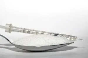
Aufgrund der wachsenden Zahl alter und sehr alter Menschen in den Industrieländern wird auch die absolute Zahl der Frakturen in den kommenden Jahrzehnten wesentlich steigen; ein Trend, der nur dann rückgängig gemacht werden kann, wenn die Prävention und die rechtzeitige Diagnose und Therapie mehr Aufmerksamkeit erhalten.

Die beiden wichtigsten zugrunde liegenden Mechanismen von Frakturen sind Osteoporose und Stürze [1]. Dabei sind Oberschenkel-Frakturen die häufigsten und kostenintensivsten, Frakturen des Beckens die tödlichsten. Die Arbeitsgruppe um Bleibler rechnet, dass in Deutschland die Zahl von Osteoporose-bedingten Frakturen von 115.248 aus dem Jahre 2010 bis zum Jahre 2050 auf 273.794 anwachsen wird. Kumuliert sind das etwa 8,1 Millionen Frakturen (78 % Frauen, 22 % Männer). Die Kosten werden von etwa 1 Milliarde in 2010 auf 6,1 Milliarden Euro in 2050 steigen. Kumuliert sind das etwa 88,5 Milliarden Euro. Der Verlust an QALYs (quality adjusted life years) liegt bei etwa 2,5 Millionen [2]. Im Jahr 2010 wurde die Zahl der Todesfälle in der Europäischen Union im Zusammenhang mit osteoporotischen Frakturen auf etwa 43.000 geschätzt [3].

Was sind Osteopenie und Osteoporose?

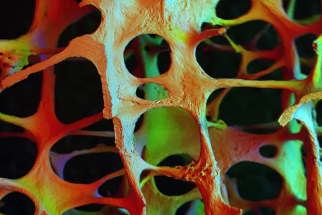
Osteopenie (Vorstufe) und Osteoporose sind systemische Erkrankung des Skeletts, die durch eine reduzierte Knochenmasse, eine Verschlechterung der Mikroarchitektur des Knochengewebes und eine erhöhte Knochenbrüchigkeit gekennzeichnet sind [4]. Diese Knochenpathologie kann in primäre oder sekundäre Formen klassifiziert werden. Die primäre Osteoporose ist durch einen progressiven, altersbedingten Mineralienverlust des Knochens gekennzeichnet, der vor allem durch Veränderungen im Sexualhormon-Haushalt beeinflusst wird.

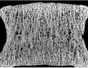
Gesunder Wirbelkörper
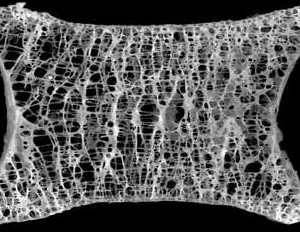
Wirbelkörper mit Osteopenie
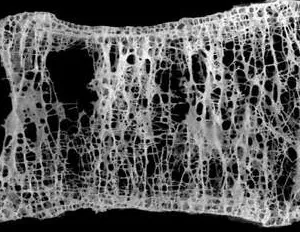
Wirbelkörper mit Osteoporose

Hier wird wiederum zwischen einer postmenopausalen (Typ I) oder einer senilen Osteoporose (Typ II) unterschieden. Die Typ-I Patienten sind Frauen, in der Regel zwischen 50 und 65 Jahren, mit Östrogenmangel und einer progressiven trabekulären Knochenresorption vor allem in Wirbelsäule und Handgelenken. Im Gegensatz dazu ist die senile Osteoporose Typ II durch einen ausgeglichenen Verlust von kortikalen und spongiösen Bereichen des Knochengewebes gekennzeichnet. Charakteristische Typ II-Osteoporose-Frakturen sind Brüche des proximalen Humerus, der Tibia, der Hüfte und des Beckens [5].

Altersspezifische Häufigkeit von Osteopenie und Osteoporose

Alter Normale BMD
Männer (%)
Normale BMD
Frauen (%)
Osteopenie
Männer (%)
Osteopenie
Frauen (%)
Osteoporose
Männer (%)
Osteoporose
Frauen (%)
65-69 20,1 22,3 65,8 60,5 14,1 17,2
70-74 22,3 15,5 64,4 62,2 13,3 22,3
75-79 20,9 12,2 59,9 57,8 19,2 30,0
80-84 19,2 11,3 58,3 54,4 22,5 34,3
85+ 12,1 6,5 51,5 48,4 36,4 45,2
Alter
(Jahre)
Normale BMD
Männer (%)
Normale BMD
Frauen (%)
65-69 20,1 22,3
70-74 22,3 15,5
75-79 20,9 12,2
80-84 19,2 11,3
85+ 12,1 6,5
Alter
(Jahre)
Osteopenie
Männer (%)
Osteopenie
Frauen (%)
65-69 65,8 60,5
70-74 64,4 62,2
75-79 59,9 57,8
80-84 58,3 54,4
85+ 51,5 48,4
Alter
(Jahre)
Osteoporose
Männer (%)
Osteoporose
Frauen (%)
65-69 14,1 17,2
70-74 13,3 22,3
75-79 19,2 30,0
80-84 22,5 34,3
85+ 36,4 45,2

Alters- und geschlechtsspezifische Verteilung von Osteoporose und Osteopenie. Abk.: BMD = Bone Mineral Density | Knochenmineraldichte. Quelle: Benzinger P, et al. BMC Geriatr. 2016; 16: 75. [1]

Osteoporose bei Männern

Osteoporose wurde lange Zeit als eine Frauen-Krankheit betrachtet, allerdings steigen alterungsbedingte Frakturen auch bei Männern an. Die genaue Zahl erkrankter Männer ist jedoch unbekannt, vermutlich aufgrund der geringeren Screening-Häufigkeit und Prüfung der Knochenmineraldichte (BMD). Je nach Land werden bisher unterschiedliche Schätzdaten angenommen: So z. B. in den USA etwa 1-2 Millionen Männer [6]. In Dänemark wurden in einer Stichprobe von 600 Männern (60 und 75 Jahre) 10,2 % mit Osteoporose diagnostiziert [7]. Insgesamt wird angenommen, dass etwa die Hälfte aller Frauen und ein Viertel aller Männer über 50 Jahre eine osteoporotische Fraktur erleiden werden [4].

Niereninsuffizienz und Sturzrisiko

Alte Menschen haben nach schweren Stürzen eine schlechte Prognose. Die Arbeitsgruppe um Bowling untersuchte, ob sich die Mortalität aufgrund von schweren Stürzen bei niereninsuffizienten Patienten (≥ 65 Jahre) erhöht. Dabei wurden die Daten von 2.590 Patienten mit Niereninsuffizienz (eGFR < 60 ml / min pro 1,73 m² oder Albumin-Kreatinin-Verhältnis ≥ 30 mg/g) analysiert. Schwere Sturz-Verletzungen wurden als sturzbedingten Fraktur, Gehirnverletzung oder Luxation definiert und die kumulative Mortalität mit einer altersentsprechenden Kontrollgruppe verglichen. Bowling et al. konnten zeigen, dass bei niereninsuffizienten Patienten die kumulative 1-Jahres-Mortalitätsrate nach einem schweren Sturz mit 21 % deutlich höher war als bei der Kontrollgruppe (5,5 %). Dabei war das Sturzrisiko vor allem mit einem erhöhten Albumin-Kreatinin-Verhältnis verbunden [8].

Hüftfrakturen mit schlechter Prognose

Die Auswirkungen von Frakturen auf das Überleben hängen von der Art der Fraktur ab. Hüftfrakturen sind am gefährlichsten, da etwa 10-20 % der Frauen mit einer Hüftfraktur innerhalb des ersten Jahres sterben. Und zwar deutlich früher als für das erreichte Alter zu erwarten wäre. Auch bei Männern ist die Sterblichkeit höher, obwohl die Sterblichkeitsraten nach Hüftfrakturen in den letzten 20 Jahren konstant geblieben zu sein scheinen. Dabei ist das Sterberisiko unmittelbar nach der Fraktur am höchsten und nimmt im Laufe der Zeit ab. Osteoporosebedingte Frakturen führen sehr häufig zu einer Beeinträchtigung der körperlichen Funktionsfähigkeit, einschließlich des Verlustes der Mobilität und der Fähigkeit, sich selbst zu versorgen. Etwa 7 % der Frauen sind bei den grundlegenden Aktivitäten des täglichen Lebens auf die Hilfe anderer angewiesen, weitere 8 % müssen in einem Pflegeheim untergebracht werden. Die größte langfristige Beeinträchtigung betrifft die Gehfähigkeit: Die Hälfte der Patienten, die vor der Fraktur gehen konnten, sind nach der Fraktur nicht mehr in der Lage, selbständig zu gehen. Darüber hinaus kann bis zu einem Drittel der Menschen, die eine Hüftfraktur erleiden, vollständig pflegebedürftig werden [4].

Weitere Risikofaktoren für Frakturen

Mehrere Faktoren tragen wesentlich zum Frakturrisiko bei, das über das durch Knochenmineraldichte-Messungen ermittelte Risiko hinausgeht. Nach den Richtlinien der "European Society for Clinical and Economic Aspects of Osteoporosis und Osteoarthritis" (ESCEO) und dem "Committees of Scientific Advisors and National Societies of the International Osteoporosis Foundation" (IOF) [9] gehören dazu Alter, Geschlecht, niedriger Body-Mass-Index, frühere Frakturen, elterliche Vorgeschichte einer Hüftfraktur, Behandlung mit Glukokortikoiden, Rauchen, Alkoholkonsum und die Ursachen für eine sekundäre Osteoporose.

Weitere Risikofaktoren, die bei der Beurteilung des Risikos von Nutzen sind, sind eine Reduktion der Körperlänge (> 4 cm) und eine pathologische Brustkyphose. Eine pathologische Kyphose der Brustwirbelsäule liegt ab einer dorsal-konvexen Krümmung der Wirbelsäule von über 40° vor [9].

Auch Knochenmarker (Serumprokollagen Typ I N-Propeptid (s-PINP) und Serum-C-terminales quervernetzendes Telopeptid des Typ I-Kollagens (s-CTX) als Marker der Knochenbildung bzw. der Knochenresorption) haben eine gewisse prognostische Bedeutung, wenn eine BMD nicht verfügbar ist [9].

Osteorenale Kommunikation

Knochen sind Zielorgane für verschiedene Hormone, wie Parathormon (PTH), Vitamin D, Calcitonin und Sexualhormone. PTH und Vitamin D beeinflussen erheblich den Serum-Calcium- sowie -Phosphat-Spiegel und damit Knochen-Bildung und -Abbau. Die Synthese und Freisetzung dieser beiden Hormone wird hauptsächlich durch den Serum-Calcium-Spiegel geregelt [10]. Bei normaler Nierenfunktion wandelt die Niere den Vitamin D-Metaboliten 25-Hydroxyvitamin D3 (25(OH)D₃) in 1,25(OH)₂D₃ (aktives Vitamin D3, Calcitriol) um. Das dabei entstehende aktive Vitamin D stimuliert die Absorption von Calcium und Phosphat im Darm sowie die Reabsorption in der Niere und die Mobilisierung aus den Knochen [11].

25(OH)D₃-Spiegel in der deutschen Bevölkerung

Alter <30 nmol/l 30-<50 nmol/l 50-<75 nmol/l ≥75 nmol/l
Frauen (18-44 Jahre) 28,9 % 27,0 % 26,6 % 17,5 %
Frauen (45-64 Jahre) 28,8 % 34,5 % 27,9 % 8,9 %
Frauen (65-79 Jahre) 32,9 % 36,9 % 24,4 % 5,7 %
Frauen (gesamt) 29,7 % 31,8 % 26,6 % 11,9 %
Männer (18-44 Jahre) 32,8 % 28,7 % 24,2 % 14,2 %
Männer (45-64 Jahre) 30,4 % 31,1 % 28,3 % 10,2 %
Männer (65-79 Jahre) 26,6 % 36,0 % 29,1 % 8,2 %
Männer (gesamt) 30,8 % 30,9 % 26,6 % 11,6 %
Gesamt-Bevölkerung 30,2 % 31,3 % 26,6 % 11,8 %

Alters- und geschlechtsspezifische Verteilung der 25(OH)D₃-Spiegel in der deutschen Bevölkerung.
Quelle: Rabenberg M, et al. BMC Public Health. 2015; 15: 641.

Parathyreoidhormon (PTH) und Niereninsuffizienz

Bei chronischer Niereninsuffizienz kommt es bereits früh zu einem Calcitriol-Defizit und einer Phosphatretention, die durch den Verlust der glomerulären Filtrationsrate (GFR) und durch einen relativen Vitamin D-Mangel bedingt sein kann. Infolge des Vitamin D-Mangels und der Hyperphosphatämie entwickelt sich eine Hypokalzämie (Serum-Kalzium unter 2,2-2,65 mmol/l). Eine Hypokalzämie signalisiert der Nebenschilddrüse PTH auszuschütten, welches wiederum die 1α-Hydroxylase des 25(OH)D₃ (25-Hydroxyvitamin D3, Cholecalciferol, Calcifediol oder Calcidiol) und damit die renale Vitamin D-Produktion stimuliert und auf diese Weise die Calciumresorption in Niere und Darm erhöht [12].

Daher steigt bereits in der Prädialyse reaktiv das PTH, weil die geschädigten Nieren nicht mehr genügend Vitamin D produzieren [13]. Darüber hinaus kann PTH den Phosphat-Efflux aus den Knochen erhöhen und stimuliert zusätzlich die Synthese des Peptidhormons Fibroblasten-Wachstumsfaktor 23 (FGF23) [14]. Ein hoher PTH-Spiegel führt ferner indirekt zur Reifung und Aktivierung der Osteoklasten und damit zu einer Calcium-Phosphat-Mobilisierung aus dem Knochengewebe, um Calciumdefizite im Blut auszugleichen [15].

Diagramm aus der Studie Movilli et al. zu Parathormon und chronischer Azidose

Gerät bei chronischer Niereninsuffizienz diese osteorenale Kommunikation aus dem Gleichgewicht sind zahlreiche therapeutische Konzepte zu bedenken und einzusetzen, Komorbiditäten wie erhöhter Blutdruck und Diabetes stringent zu behandeln und ein engmaschiges Screening durchzuführen, um eine Progression der Erkrankungen zu vermeiden [16].

Fibroblasten-Wachstumsfaktor 23 (FGF23)

Mikroskopische Aufnahme von Eierstockkrebs
Konfokale Mikroskopie eines Fibroblasten mit Zellkern (blau), Mitochondrien (rot) und Aktinzytoskelett (grün). Quelle: National Cancer Institute. Download: Bild als Druckversion / Wallpaper in neuem Fenster öffnen. (3.680 kb)
FGF23 wird überwiegend von Osteozyten gebildet und inhibiert die renale tubuläre Reabsorption von Phosphat und erhöht damit die Urin-Phosphat-Ausscheidung. FGF23 reduziert die renale Phosphatrückresorption, indem es die apikale Expression des Natrium-abhängigen Phosphat-Kotransporters-IIa (NaPi-IIa) in der Bürstenmembran des proximal renalen Tublus hemmt [17]. Die Expression und Aktivität dieser Transporter wird durch PTH, Calcitriol, Glukokortikoide, Phosphat und durch FGF23 reguliert [18].

Erhöhte Phosphatspiegel stimulieren die Ausschüttung von FGF23 aus den Osteozyten und Osteoblasten, während erniedrigte Phosphatspiegel zu reduzierten FGF23-Werten führen. 1,25(OH)2D3 (aktives Vitamin D) stimuliert ebenfalls eine Ausschüttung von FGF23. Ferner hemmt FGF23 die Synthese des aktiven Vitamin D, indem es die 1αHydroxylase inhibiert. Dadurch sinkt der Calcitriolspiegel. Reduzierte Calcitriolspiegel wiederum verringern die intestinale Phosphat- und Calciumresorption [19]. Die Konzentrationen von FGF23 korrelieren ferner positiv mit der diätetischen Calciumzufuhr (p = 0.01), allerdings nicht nach akut erhöhter Calciumzufuhr [20].

Modeling und Remodeling von Knochen

Der Knochen adaptiert seine mechanische Funktion, das heißt seine Geometrie und damit die Knochenfestigkeit, ein Leben lang auf die täglichen Anforderungen. Das Modell Mechanostat von Harald Frost beschreibt die Dynamik des Knochenumbaus. Demnach wird Knochenwachstum und Knochenabbau durch die maximale elastische Verformung des Knochens bestimmt. Dabei sind die auftretenden kurzzeitigen Maximalkräfte entscheidend für den Umbau.

Diese Tatsache hat gerade auch für Knochenschwund therapeutische Konsequenzen: Geeignetes Training mit mechanischen Spitzenkräften auf den Knochen kann das Knochenwachstum stimulieren, die Knochenfestigkeit steigern und so einer Osteoporose entgegenwirken. Für den Knochenaufbau förderlich sind dynamische Übungen mit Krafteinsatz, ein Ausdauer-orientiertes Training (z.B. Laufen, Radfahren, Schwimmen) hat keinen (oder wenig) Einfluss auf die Knochenbildung, da die auftretenden Kraftspitzen zu gering sind. Bewährt hat sich vor allem das Vibrationstraining (auch Whole Body Vibration (WBV), Beschleunigungstraining, Schwingungstraining, Mechanostimulation oder stochastisches Resonanztraining).

Frost hat vier Bereiche der Knochenverformung, gemessen in μStrain, definiert:

Disuse: Strain < circa 800 μStrain: Remodeling (Knochenumbau und Knochenreparatur) findet statt, Knochenmasse und Knochenfestigkeit wird abgebaut.

Adapted State: Strain zwischen ca. 800 und 1.500 μStrain: Remodeling findet statt, Knochenmasse und Knochenfestigkeit bleiben unverändert.

Overload: Strain > ca. 1.500 μStrain: Modeling (Knochenaufbau) findet statt, Knochenmasse und Knochenfestigkeit werden vergrößert.

Fracture: Strain > ca. 15.000 μStrain: Bruchgrenze, der Knochen bricht.

1000 μStrain = entsprechen 0,1 % Längenänderung

Zu berücksichtigen ist hierbei, dass die Festigkeit des Knochens stark von der Geometrie und von der Richtung der Krafteinleitung abhängig ist. Die Tibia beispielsweise hat in axialer Richtung etwa eine Bruchgrenze vom 50- bis 60-fachen Körpergewicht. Senkrecht zu dieser Achse liegt die Bruchgrenze jedoch um den Faktor 10 oder mehr niedriger.

α-Klotho

Als weiterer Biomarker des Phosphat- und Vitamin D-Stoffwechsels bei frühen chronischen Nierenerkrankungen gilt das α-Klotho [21]. Es wird vorwiegend in der Niere als membranständiges Protein (TM-Klotho) und als zirkulierende lösliche Variante (s-Klotho) produziert. Eine tierexperimentelle Studie konnte in vivo durch Micropunktion und in vitro an proximalen Tubuluszelllinien zeigen, dass α-Klotho in den Nieren sezerniert wird und transcytotisch von der basalen zur apikalen Seite der proximalen Tubuluszellen migriert [22]. Daraus schließen die Autoren, dass die Niere zwei Aufgaben in der Aufrechterhaltung der α-Klotho-Homöostase hat: Produktion und Freisetzung sowie Clearing aus dem Blut in den Urin. α-Klotho fungiert als Cofaktor für den FGF23-Rezeptor und erhöht die Affinität für die FGF23-Bindung am Rezeptor.

Therapie der Osteoporose

Lebensstil-Änderungen und Ernährungsmaßnahmen bei Osteoporose

  1. Tägliche Kalziumzufuhr zwischen 800 und 1200 mg und ausreichend Nahrungsprotein, idealerweise über Milchprodukte.
  2. Bei postmenopausalen Frauen mit erhöhtem Frakturrisiko sollte eine tägliche Dosis von 800 IE Cholecalciferol empfohlen werden.
  3. Eine Kalziumergänzung ist angebracht, wenn die ernährungsbedingte Aufnahme unter 800 mg/Tag liegt und eine Vitamin-D-Supplementierung bei Patienten in Betracht gezogen wird, die ein Risiko für eine Vitamin-D-Insuffizienz haben oder Anzeichen einer solchen aufweisen. Achtung: Durch die Gabe von Kalzium in hohen Mengen kann die Ausscheidung mit dem Urin (Hyperkalziurie) ansteigen. Folge können Nierensteine oder eine Nephrokalzinose sein.
  4. Regelmäßige, auf die Bedürfnisse und Fähigkeiten der einzelnen Patienten zugeschnittene körperliche Belastung.
  5. Bei Personen mit erhöhtem Frakturrisiko sollte eine Sturz-Anamnese erstellt werden, wobei bei erhöhtem Risiko eine weitere Beurteilung und geeignete Maßnahmen durchgeführt werden sollten.

Quelle: Kanis, JA et al. Calcif Tissue Int. 2019 Mar; 104(3): 235–238. [9]

Pharmakologische Interventionen bei postmenopausalen Frauen

  1. Die oralen Bisphosphonate (Alendronat, Risedronat und Ibandronat) können in der Mehrzahl der Fälle als Erstbehandlung eingesetzt werden. Bei Frauen, die orale Bisphosphonate nicht vertragen (oder bei Frauen, bei denen sie kontraindiziert sind), stellen intravenöse Bisphosphonate oder Denosumab die am besten geeigneten Alternativen dar, wobei Raloxifen oder die Hormontherapie der Wechseljahre als zusätzliche Optionen zur Verfügung stehen. Teriparatid wird bevorzugt für Patienten mit hohem Frakturrisiko empfohlen.
  2. Die Therapie mit Bisphosphonat sollte nach 3 - 5 Jahren überprüft werden. Das Frakturrisiko sollte nach jedem Knochenbruch neu bewertet werden. Das Risiko von Wirbelkörpern-Frakturen steigt bei Patienten, die die Behandlung abbrechen.
  3. Das Absetzen einer Denosumab-Therapie ist mit einem Anstieg der Wirbelkörper-Frakturrate verbunden. Eine Bisphosphonat-Therapie kann nach Absetzen von Denosumab in Betracht gezogen werden.
  4. Es gibt kaum Anhaltspunkte für eine Entscheidungsfindung, die über 10 Jahre Behandlung hinausgeht. Daher sollten die medikamentöse Therapie und andere Behandlungsoptionen sowie der Therapieerfolg bei Osteoporose-Patienten auf individueller Basis regelmäßig geprüft werden.

Quelle: Kanis, JA et al. Calcif Tissue Int. 2019 Mar; 104(3): 235–238. [9]

Pharmakologische Interventionen bei Niereninsuffizienz und chronischer Azidose

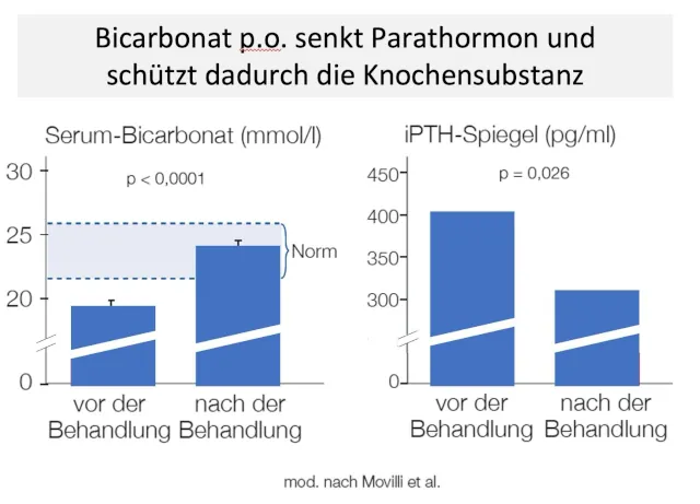
Plasma-Bikarbonat ist mit der Knochenmineraldichte und der Knochenabbaurate (Knochenschwund) assoziiert. Das zeigt, dass der systemische Säure-Basen-Status eine wichtige Determinante der Skelett-Gesundheit während des Alterns ist [23, 24]:

Das Säure-Basen-Gleichgewicht wird wesentlich durch die Lunge und Nieren reguliert. Ferner neutralisieren körpereigene Puffersubstanzen, wie beispielsweise Bicarbonat, im Stoffwechsel anfallende Säuren und sind auf diese Weise an der Regulierung des pH-Wertes in Blut und Geweben beteiligt. Bei nicht normaler Regulierung verursacht die Anhäufung von Wasserstoffionen (H+) eine metabolische Azidose. Diese stört den Stoffwechsel von Knochen und Muskeln und kann darüber hinaus zu Anomalien bei der Freisetzung und Funktion verschiedener Hormone führen, darunter Wachstumshormone, IGF-1, Insulin, Glukokortikoide, Schilddrüsenhormone, Nebenschilddrüsenhormon und Vitamin D. Klinische Folgen dieser anormalen Stoffwechselreaktionen sind Wachstumsstörungen bei Säuglingen und Kindern sowie der Verlust von Knochen- und Muskelmasse bei Erwachsenen [25].

Die chronische metabolische Azidose und ihre Folgen können durch eine Behandlung mit Natriumhydrogenkarbonat (NaHCO₃ / kurz: Bikarbonat) korrigiert werden [25]. Dabei scheint Natriumhydrogenkarbonat die beste Alternative zu anderen Azidosetherapeutika zu sein, weil z. B.Patienten mit Nierensteinen andere alkalisierende Mittel wie z. B. Kaliumcitrat wegen der Nebenwirkungen (Hyperkaliämie und Magendarmstörungen) nicht vertragen können. Natriumbikarbonat erwies sich als ähnlich wirksam, kostengünstiger und nebenwirkungsärmer [26]. Das gilt insbesondere für magensaftresistente Präparate [27]. U. a. wegen gastrointestinaler Nebenwirkungen und besserer Bioverfügbarkeit empfiehlt eine interdisziplinäre Kommission, Bikarbonat ausschließlich in magensaftresistenter Galenik einzunehmen. Pharmakologisch sinnvoll sind dabei allerdings nur Präparate mit einer Galenik, die eine schnelle Freisetzung des Wirkstoffes im oberen Dünndarm gewährleisten, während eine retardierte langsame Freisetzung (slow release) unerwünscht auch untere Darmabschnitte alkalisieren kann.

Fazit: Osteoporose ist behandelbar

In der ärztlichen Praxis kommt Osteoporose relativ häufig bei postmenopausalen Frauen aber auch bei älteren Männern vor. Die Pathogenese ist vielfältig, von hormonellen Störungen, Nierenschwäche bis hin zu einer chronischen metabolischen Azidose. Die Therapie beinhaltet neben Änderungen des Lebenstils auch medikamentöse Interventionen. Dabei können auch Nahrungsergänzungsmittel sinnvoll sein (Kalzium, Vitamin D). Wenn eine Nierenschwäche vorliegt, ist aufgrund der mangelnden Ausscheidung von Säuren die chronische Azidose eine häufige Komplikation, die mit Azidosetherapeutike, wie Natriumbikarbonat, behandelt werden muss, um die Progression von Osteoporose, Niereninsuffizienz und Azidose zu stoppen oder zumindest abzumildern.

Literatur

Abstract: The Impact of Preventive Measures on the Burden of Femoral Fractures - A Modelling Approach to Estimating the Impact of Fall Prevention Exercises and Oral Bisphosphonate Treatment for the Years 2014 and 2025

Background: Due to the demographic transition with a growing number of old and oldest-old persons the absolute number of fragility fractures is expected to increase in industrialized countries unless effective preventive efforts are intensified. The main causes leading to fractures are osteoporosis and falls. The aim of this study is to develop population based models of the potential impact of fall-prevention exercise and oral bisphosphonates over the coming decade.

Methods: The German federal state of Bavaria served as the model population. Model interventions were limited to community-dwelling persons aged 65 years and older. Models are based on fall-prevention exercise being offered to all persons aged 70 to 89 years and oral bisphosphonate treatment offered to all persons with osteoporosis as defined by a T-score of ≤ − 2.5. Treatment effect sizes are estimated from meta-analyses. Reduction in all femoral fractures in the population of community-dwelling persons aged 65 years and older is the outcome of interest. A spreadsheet-based modelling approach was used for prediction.

Results: In 2014, reduction of femoral fractures by 10 % required 21 % of all community-dwelling persons aged 70–89 to participate in fall-prevention exercise, or 37 % of those with osteoporosis to receive oral bisphosphonates. Without intervention, demographic changes will result in a 24 % increase in femoral fractures by 2025. To lower the increase of fractures between 2014 and 2025 to 10 %, fall-prevention-exercise participation rate needs to be 25 % and bisphosphonate treatment rates 41 %, whereas to hold the 2025 rates flat at 2014 rates require 43 % fall-prevention-exercises participation, and is not achievable using oral bisphosphonates.

Conclusions: Unrealistic high treatment and participation rates of the two analysed measures are needed to achieve substantial effects on the expected burden of femoral fractures at present and in the future.

Keywords: Epidemiology; Falls; Femoral fractures; Osteoporosis; Prevention.

Published by BMC Geriatrics / Part of Springer Nature | DOI: 10.1186/s12877-016-0247-9


zur Originalarbeit nach oben  

Abstract: The Health Burden and Costs of Incident Fractures Attributable to Osteoporosis From 2010 to 2050 in Germany -- a Demographic Simulation Model

To predict the burden of incident osteoporosis attributable fractures (OAF) in Germany, an economic simulation model was built. The burden of OAF will sharply increase until 2050. Future demand for hospital and long-term care can be expected to substantially rise and should be considered in future healthcare planning.

Introduction: The aim of this study was to develop an innovative simulation model to predict the burden of incident OAF occurring in the German population, aged >50, in the time period of 2010 to 2050.

Methods: A Markov state transition model based on five fracture states was developed to estimate costs and loss of quality adjusted life years (QALYs). Demographic change was modelled using individual generation life tables. Direct (inpatient, outpatient, long-term care) and indirect fracture costs attributable to osteoporosis were estimated by comparing Markov cohorts with and without osteoporosis.

Results: The number of OAF will rise from 115,248 in 2010 to 273,794 in 2050, cumulating to approximately 8.1 million fractures (78 % women, 22 % men) during the period between 2010 and 2050. Total undiscounted incident OAF costs will increase from around 1.0 billion Euros in 2010 to 6.1 billion Euros in 2050. Discounted (3 %) cumulated costs from 2010 to 2050 will amount to 88.5 billion Euros (168.5 undiscounted), with 76 % being direct and 24 % indirect costs. The discounted (undiscounted) cumulated loss of QALYs will amount to 2.5 (4.9) million.

Conclusions: We found that incident OAF costs will sharply increase until the year 2050. As a consequence, a growing demand for long-term care as well as hospital care can be expected and should be considered in future healthcare planning. To support decision makers in managing the future burden of OAF, our model allows to economically evaluate population- and risk group-based interventions for fracture prevention in Germany.

Keywords: Cost-of-illness, Demographic change, Germany, Healthcare planning, Markov model, Osteoporosis attributable fractures

Published by Springer Link | DOI: 10.1007/s00198-012-2020-z


zur Originalarbeit nach oben  

Abstract: European guidance for the diagnosis and management of osteoporosis in postmenopausal women

Guidance is provided in a European setting on the assessment and treatment of postmenopausal women at risk of fractures due to osteoporosis.

Introduction: The International Osteoporosis Foundation and European Society for Clinical and Economic Aspects of Osteoporosis and Osteoarthritis published guidance for the diagnosis and management of osteoporosis in 2008. This manuscript updates these in a European setting.

Methods: Systematic literature reviews.

Results: The following areas are reviewed: the role of bone mineral density measurement for the diagnosis of osteoporosis and assessment of fracture risk, general and pharmacological management of osteoporosis, monitoring of treatment, assessment of fracture risk, case finding strategies, investigation of patients and health economics of treatment.

Conclusions: A platform is provided on which specific guidelines can be developed for national use.

Keywords: Bone mineral density, Diagnosis of osteoporosis, Fracture risk assessment, FRAX, Health economics, Treatment of osteoporosis

Published by Springer Link | DOI: 10.1007/s00198-012-2074-y


zur Originalarbeit nach oben  

Abstract: Major Osteoporotic Fragility Fractures: Risk Factor Updates and Societal Impact

Osteoporosis is a silent disease without any evidence of disease until a fracture occurs. Approximately 200 million people in the world are affected by osteoporosis and 8.9 million fractures occur each year worldwide. Fractures of the hip are a major public health burden, by means of both social cost and health condition of the elderly because these fractures are one of the main causes of morbidity, impairment, decreased quality of life and mortality in women and men. The aim of this review is to analyze the most important factors related to the enormous impact of osteoporotic fractures on population. Among the most common risk factors, low body mass index; history of fragility fracture, environmental risk, early menopause, smoking, lack of vitamin D, endocrine disorders (for example insulin-dependent diabetes mellitus), use of glucocorticoids, excessive alcohol intake, immobility and others represented the main clinical risk factors associated with augmented risk of fragility fracture. The increasing trend of osteoporosis is accompanied by an underutilization of the available preventive strategies and only a small number of patients at high fracture risk are recognized and successively referred for therapy. This report provides analytic evidences to assess the best practices in osteoporosis management and indications for the adoption of a correct healthcare strategy to significantly reduce the osteoporosis burden. Early diagnosis is the key to resize the impact of osteoporosis on healthcare system. In this context, attention must be focused on the identification of high fracture risk among osteoporotic patients. It is necessary to increase national awareness campaigns across countries in order to reduce the osteoporotic fractures incidence.

Keywords: Fracture prevention; Fracture risk; Fragility fracture; Hip fracture; Osteoporosis.

Published by Baishideng Publishing Group | DOI: 10.5312/wjo.v7.i3.171


zur Originalarbeit nach oben  

Abstract: Radiology of Osteoporosis

Bone densitometric studies have shown that osteoporosis is a result of prolonged, slow bone loss and that the pattern of loss is different for trabecular and cortical bone. Structurally-insufficient osteoporotic bone is predisposed to fractures. Among the clinically manifest osteoporotic fractures, distal radius leads the list, followed by hip, spine, and proximal humerus. This article examines the use of conventional radiography as well as other imaging-based modalities for the evaluation of osteoporosis and associated fractures in the axial and appendicular skeleton.

Keywords: Bone densitometric, osteoporosis, conventional radiography, fractures

Published by Elsevier | DOI: 10.1016/j.rcl.2010.02.016


zur Originalarbeit nach oben  

Abstract: The Clinical Epidemiology of Male Osteoporosis: A Review of the Recent Literature

Osteoporosis, a musculoskeletal disease characterized by decreased bone mineral density (BMD) and an increased risk of fragility fractures, is now recognized as an important public health problem in men. Osteoporotic fractures, particularly of the hip, result in significant morbidity and mortality in men and lead to considerable societal costs. Many national and international organizations now address screening and treatment for men in their osteoporosis clinical guidelines. However, male osteoporosis remains largely underdiagnosed and undertreated. The objective of this paper is to provide an overview of recent findings in male osteoporosis, including pathophysiology, epidemiology, and incidence and burden of fracture, and discuss current knowledge about the evaluation and treatment of osteoporosis in males. In particular, clinical practice guidelines, fracture risk assessment, and evidence of treatment effectiveness in men are addressed.

Keywords: diagnosis; fracture risk; guidelines; screening; treatment.

Published by Dovepress | DOI: 10.2147/CLEP.S40966.


zur Originalarbeit nach oben  

Abstract: Osteoporosis and Vertebral Fractures in Men Aged 60-74 Years

Background: Limited information on the prevalence of osteoporosis and VFxs in men in high-risk populations is available. The choice of reference values for dual X-ray absorptiometry (DXA) is debated. We evaluated the prevalence of osteoporosis and vertebral deformities in a population-based sample of men.

Methods: Bone mineral density (BMD) was measured and vertebral deformities assessed using DXA and VFx assessment (VFA), respectively, in a random sample of 600 Danish men aged 60-74 years. Osteoporosis was defined as a T-score of -2.5 or less.

Results: The study population was comparable with the background population with regard to age, body mass index and co-morbidity. Osteoporosis was diagnosed in less than 1% of the participants at inclusion. Using Danish and NHANES III reference data, 10.2 and 11.5% of the study population had osteoporosis, respectively. In all, 6.3% participants had at least one VFx. BMD was significantly lower in participants with vertebral deformities, but only 24% of these cases had osteoporosis.

Conclusions: Osteoporosis and VFxs are prevalent in men aged 60-74 years. Although the majority of deformities were present in individuals without osteoporosis, BMD was lower in patients with VFxs at all sites investigated. Male osteoporosis was markedly underdiagnosed.

Keywords: men, osteoporosis, vertebral fractures, population based, elderly

Published by Oxford Academic | DOI: 10.1093/ageing/afr170.


zur Originalarbeit nach oben  

Abstract: Association of Reduced eGFR and Albuminuria With Serious Fall Injuries Among Older Adults

Background: Falls are common and associated with adverse outcomes in patients on dialysis. Limited data are available in earlier stages of CKD.

Design, setting, participants, & measurements: We analyzed data from 8744 Reasons for Geographic and Racial Differences in Stroke Study participants ≥65 years old with Medicare fee for service coverage. Serious fall injuries were defined as a fall-related fracture, brain injury, or joint dislocation using Medicare claims. Hazard ratios (HRs) for serious fall injuries were calculated by eGFR and albumin-to-creatinine ratio (ACR). Among 2590 participants with CKD (eGFR<60 ml/min per 1.73 m(2) or ACR≥30 mg/g), cumulative mortality after a serious fall injury compared with age-matched controls without a fall injury was calculated.

Results: Overall, 1103 (12.6%) participants had a serious fall injury over 9.9 years of follow-up. The incidence rates per 1000 person-years of serious fall injuries were 21.7 (95% confidence interval [95% CI], 20.3 to 23.2), 26.6 (95% CI, 22.6 to 31.3), and 38.3 (95% CI, 31.2 to 47.0) at eGFR levels ≥60, 45-59, and <45 ml/min per 1.73 m(2), respectively, and 21.3 (95% CI, 20.0 to 22.8), 31.7 (95% CI, 27.5 to 36.5), and 42.2 (95% CI, 31.3 to 56.9) at ACR levels <30, 30-299, and ≥300 mg/g, respectively. Multivariable adjusted HRs for serious fall injuries were 0.91 (95% CI, 0.76 to 1.09) and 1.09 (95% CI, 0.86 to 1.37) for eGFR=45-59 and <45 ml/min per 1.73 m(2), respectively, versus eGFR≥60 ml/min per 1.73 m(2) and 1.31 (95% CI, 1.11 to 1.54) and 1.81 (95% CI, 1.30 to 2.50) for ACR=30-299 and ≥300 mg/g, respectively, versus ACR<30 mg/g. Among participants with CKD, cumulative 1-year mortality rates among patients with a serious fall and age-matched controls were 21.0% and 5.5%, respectively.

Conclusions: Elevated ACR but not lower eGFR was associated with serious fall injuries. Evaluation for fall risk factors and fall prevention strategies should be considered for older adults with elevated ACR.

Keywords: Adult; Brain Injuries; CKD; Follow-Up Studies; Fractures, Bone; Humans; Renal Insufficiency, Chronic; albuminuria; falls; geriatric nephrology.

Published by American Society of Nephrology | DOI: 10.2215/CJN.11111015


zur Originalarbeit nach oben  

Abstract: Executive summary of the European guidance for the diagnosis and management of osteoporosis in postmenopausal women

A guidance on the assessment and treatment of postmenopausal women at risk from fractures due to osteoporosis was recently published in Osteoporosis International as a joint effort of the International Osteoporosis Foundation and European Society for Clinical and Economic Aspects of Osteoporosis and Osteoarthritis (1) This manuscript updates the previous guidelines document, published in 2013 (2) and is written in a European perspective. The present article reports and summarizes the main recommendations included in this 2018 guidance document.

Keywords: Bone mineral density, Diagnosis of osteoporosis, Fracture risk assessment, FRAX, Health economics, Treatment of osteoporosis

Published by Europe PMC Funders Group | DOI: 10.1007/s00223-018-00512-x


zur Originalarbeit nach oben  

Abstract: Calcium and Phosphorus Homeostasis

Calcium and phosphorus homeostasis relies on a complex, tightly regulated system involving many ions and hormones. The regulation of calcium and phosphorus is controlled by the actions of these ions and hormones on the intestine, kidneys and bone. Disturbances in the serum level of calcium and/or phosphorus can lead to significant pathology, including kidney stones and bone disease. In addition to parathyroid hormone and vitamin D, recently identified factors such as fibroblast growth factors and klotho play an important role in maintaining mineral ion homeostasis. The identification of subfamily V transient receptor potential cation channels (TRPV channels), Na/P(i) cotransporters, the vitamin D receptor and the calcium-sensing receptor have further advanced our understanding of this complex physiology. In this review we discuss the current understanding of the relationships between the ions, hormones, and transporters that maintain calcium and phosphorus homeostasis.

Keywords: Calcium, phosphorus, homeostasis, kidney stones, bone disease, parathyroid hormone, vitamin D, fibroblast growth factors, klotho

Published by Karger | DOI: 10.1159/000209740


zur Originalarbeit nach oben  

Abstract: Fibroblast Growth factor-23 in Early Chronic Kidney Disease: Additional Support in Favor of a Phosphate-Centric Paradigm for the Pathogenesis of Secondary Hyperparathyroidism

Background: The discovery of fibroblast growth factor-23 (FGF-23) and the elucidation of its function as a phosphaturic and 1,25(OH)2VitD counter-regulatory hormone provides a new conceptual framework for the understanding of the pathogenesis of secondary hyperparathyroidism. This study aims to elucidate the complex associations between FGF-23, parathyroid hormone (PTH), 1,25(OH)2D, and phosphate in patients with early-stage chronic kidney disease (CKD) and to provide clinical evidence in favor of the new phosphate-centric paradigm for the pathogenesis of secondary hyperparathyroidism.

Design, setting, participants, & measurements: Serum biointact PTH and FGF-23, 25(OH)D, 1,25(OH)2D, calcium, phosphate, 24-hour urine excretion of phosphate and calcium, and urinary fractional excretion of phosphate were determined in a cross-sectional study including 125 patients with CKD stages 1 to 3.

Results: Serum phosphate levels showed an inverse association with estimated GFR (eGFR), but were within the normal range in all but one patient. FGF-23 and PTH were inversely associated with eGFR, even in the subgroup of patients with CKD stages 1 and 2. High FGF-23 levels were significantly more prevalent than high PTH levels. The urinary fractional excretion of phosphate was highest in patients with both a high serum FGF-23 and PTH level. Increased FGF-23 and phosphate and decreased 25(OH)D were independently associated with decreased 1,25(OH)2D.

Conclusions: Our data are in favor of the new paradigm for the pathogenesis of secondary hyperparathyroidism according to which a reduced phosphate excretion capacity is the principal abnormality that initiates secondary hyperparathyroidism.

Keywords:

Published by American Society of Nephrology | DOI: 10.2215/CJN.08241109


zur Originalarbeit nach oben  

Abstract: Dysregulation of phosphate metabolism and conditions associated with phosphate toxicity

Phosphate homeostasis is coordinated and regulated by complex cross-organ talk through delicate hormonal networks. Parathyroid hormone (PTH), secreted in response to low serum calcium, has an important role in maintaining phosphate homeostasis by influencing renal synthesis of 1,25-dihydroxyvitamin D, thereby increasing intestinal phosphate absorption. Moreover, PTH can increase phosphate efflux from bone and contribute to renal phosphate homeostasis through phosphaturic effects. In addition, PTH can induce skeletal synthesis of another potent phosphaturic hormone, fibroblast growth factor 23 (FGF23), which is able to inhibit renal tubular phosphate reabsorption, thereby increasing urinary phosphate excretion. FGF23 can also fine-tune vitamin D homeostasis by suppressing renal expression of 1-alpha hydroxylase (1α(OH)ase). This review briefly discusses how FGF23, by forming a bone–kidney axis, regulates phosphate homeostasis, and how its dysregulation can lead to phosphate toxicity that induces widespread tissue injury. We also provide evidence to explain how phosphate toxicity related to dietary phosphorus overload may facilitate incidence of noncommunicable diseases including kidney disease, cardiovascular disease, cancers and skeletal disorders.

Keywords: Phosphate homeostasis, Parathyroid hormone, serum calcium, 1,25-dihydroxyvitamin D, fibroblast growth factor 23, klotho

Published by Nature Publishing Group | DOI: 10.1038/bonekey.2015.74


zur Originalarbeit nach oben  

Abstract: Osteo-renal Regulation of Systemic Phosphate Metabolism

Impaired kidney function and subsequent skeletal responses play a critical role in disrupting phosphate balance in chronic kidney disease (CKD) patients with mineral and bone disorder (CKD-MBD). In patients with CKD-MBD, the inability of the kidney to maintain normal mineral ion balance affects bone remodeling to induce skeletal fracture and extraskeletal vascular calcification. In physiological conditions, bone-derived fibroblast growth factor 23 (FGF23) acts on the kidney to reduce serum phosphate and 1,25-dihydroxyvitamin D levels. In humans, increased bioactivity of FGF23 leads to increased urinary phosphate excretion, which induces hypophosphatemic diseases (e.g., rickets/osteomalacia). However, reduced FGF23 activity is associated with hyperphosphatemic diseases (e.g., tumoral calcinosis). In patients with CKD, high serum levels of FGF23 fail to reduce serum phosphate levels and lead to numerous complications, including vascular calcification, one of the important determinants of mortality of CKD-MBD patients. Of particular significance, molecular, biochemical and morphological changes in patients with CKD-MBD are mostly due to osteo-renal dysregulation of mineral ion metabolism. Furthermore, hyperphosphatemia can partly contribute to the development of secondary hyperparathyroidism in patients with CKD-MBD. Relatively new pharmacological agents including sevelamer hydrochloride, calcitriol analogs and cinacalcet hydrochloride are used either alone, or in combination, to minimize hyperphosphatemia and hyperparathyroidism associated complications to improve morbidity and mortality of CKD-MBD patients. This article will briefly summarize how osteo-renal miscommunication can induce phosphate toxicity, resulting in extensive tissue injuries.

Keywords: Klotho; fibroblast growth factor 23; vitamin D; parathyroid hormone; chronic kidney disease

Published by Wiley Periodicals, Inc. | DOI: 10.1002/iub.437


zur Originalarbeit nach oben  

Abstract: Regulatory Mechanism of Circulating Inorganic Phosphate

Circulating level of phosphate is altered by age and diet, and is also controlled by several hormones such as parathyroid hormone(PTH), 1,25-dihydroxyvitamin D[1,25(OH)2D]and fibroblast growth factor 23(FGF23). The main function of PTH and 1,25(OH)2D is maintaining calcium homeostasis, while FGF23 plays a central role in phosphate metabolism. PTH suppresses phosphate reabsorption in the proximal tubules to increase the renal phosphate wasting, while 1,25(OH)2D facilitates the intestinal phosphate absorption. FGF23 increases the renal phosphate wasting and reduces the production of 1,25(OH)2D. Of note, these hormones mutually regulate one another. The production of FGF23 is also regulated by various local factors. The mechanism for sensing the phosphate availability still remains unknown, and further investigation is required.

Keywords:

Abstract published by PubMed.gov | PMID: 26813498


Kein Link [Artikel auf Japanisch] nach oben  

Abstract: Lifelong Challenge of Calcium Homeostasis in Male Mice Lacking TRPV5 Leads to Changes in Bone and Calcium Metabolism

Trpv5 plays an important role in calcium (Ca2+) homeostasis, among others by mediating renal calcium reabsorption. Accordingly, Trpv5 deficiency strongly stresses Ca2+ homeostasis in order to maintain stable serum Ca2+. We addressed the impact of lifelong challenge of calcium homeostasis on the bone phenotype of these mice.Aging significantly increased serum 1,25(OH)2D3 and PTH levels in both genotypes but they were more elevated in Trpv5-/- mice, whereas serum Ca2+ was not affected by age or genotype. Age-related changes in trabecular and cortical bone mass were accelerated in Trpv5-/- mice, including reduced trabecular and cortical bone thickness as well as reduced bone mineralization. No effect of Trpv5 deficiency on bone strength was observed. In 78-week-old mice no differences were observed between the genotypes regarding urinary deoxypyridinoline, osteoclast number, differentiation and activity as well as osteoclast precursor numbers, as assessed by flow cytometry.In conclusion, life-long challenge of Ca2+ homeostasis present in Trpv5-/- mice causes accelerated bone aging and a low cortical and trabecular bone mass phenotype. The phenotype of the Trpv5-/- mice suggests that maintenance of adequate circulatory Ca2+ levels in patients with disturbances in Ca2+ homeostasis should be a priority in order to prevent bone loss at older age.

Keywords: Gerotarget; TRPV5; aging; bone resorption; calcium homeostasis; mineralization.

Published by Oncotarget | DOI: 10.18632/oncotarget.8779


zur Originalarbeit nach oben  

Abstract: Stop Chronic Kidney Disease Progression: Time Is Approaching

Progression of chronic kidney disease (CKD) is inevitable. However, the last decade has witnessed tremendous achievements in this field. Today we are optimistic; the dream of withholding this progression is about to be realistic. The recent discoveries in the field of CKD management involved most of the individual diseases leading the patients to end-stage renal disease. Most of these advances involved patients suffering diabetic kidney disease, chronic glomerulonephritis, polycystic kidney disease, renal amyloidosis and chronic tubulointerstitial disease. The chronic systemic inflammatory status and increased oxidative stress were also investigated. This inflammatory status influences the anti-senescence Klotho gene expression. The role of Klotho in CKD progression together with its therapeutic value are explored. The role of gut as a major source of inflammation, the pathogenesis of intestinal mucosal barrier damage, the role of intestinal alkaline phosphatase and the dietary and therapeutic implications add a novel therapeutic tool to delay CKD progression.

Keywords: Amyloidosis; Chronic kidney disease; Diabetic nephropathy; Klotho; Micro RNA; Progression.

Published by Baishideng Publishing Group | DOI: 10.5527/wjn.v5.i3.258


zur Originalarbeit nach oben  

Abstract: Structural Fold and Binding Sites of the Human Na⁺-phosphate Cotransporter NaPi-II

Phosphate plays essential biological roles and its plasma level in humans requires tight control to avoid bone loss (insufficiency) or vascular calcification (excess). Intestinal absorption and renal reabsorption of phosphate are mediated by members of the SLC34 family of sodium-coupled transporters (NaPi-IIa,b,c) whose membrane expression is regulated by various hormones, circulating proteins, and phosphate itself. Consequently, NaPi-II proteins are also potentially important pharmaceutical targets for controlling phosphate levels. Their crucial role in Pi homeostasis is underscored by pathologies resulting from naturally occurring SLC34 mutations and SLC34 knockout animals. SLC34 isoforms have been extensively studied with respect to transport mechanism and structure-function relationships; however, the three-dimensional structure is unknown. All SLC34 transporters share a duplicated motif comprising a glutamine followed by a stretch of threonine or serine residues, suggesting the presence of structural repeats as found in other transporter families. Nevertheless, standard bioinformatic approaches fail to clearly identify a suitable template for molecular modeling. Here, we used hydrophobicity profiles and hidden Markov models to define a structural repeat common to all SLC34 isoforms. Similar approaches identify a relationship with the core regions in a crystal structure of Vibrio cholerae Na(+)-dicarboxylate transporter VcINDY, from which we generated a homology model of human NaPi-IIa. The aforementioned SLC34 motifs in each repeat localize to the center of the model, and were predicted to form Na(+) and Pi coordination sites. Functional relevance of key amino acids was confirmed by biochemical and electrophysiological analysis of expressed, mutated transporters. Moreover, the validity of the predicted architecture is corroborated by extensive published structure-function studies. The model provides key information for elucidating the transport mechanism and predicts candidate substrate binding sites.

Keywords: Phosphate; bone loss; vascular calcification; sodium-coupled transporter;

Published by Elsevier Inc. / CellPress Open Access | DOI: 10.1016/j.bpj.2014.01.043.


zur Originalarbeit nach oben  

Abstract: Vitamin D and Type II Sodium-Dependent Phosphate Cotransporters

The type II sodium-dependent Pi (NaPi) cotransporters (NaPi-IIa, NaPi-IIb and NaPi-IIc) contribute to renal and intestinal Pi absorption. 1,25-Dihydroxyvitamin D [1,25(OH)2D3] is an important factor for NaPi-II transporters in the small intestine and kidney. In a previous study, low levels of 1,25(OH)2D3 appeared to suppress the expression of renal NaPi cotransporters. We identified a functional vitamin D receptor-responsive element in the human NaPi-IIa and NaPi-IIc genes in renal epithelial cells. In an analysis of vitamin D receptor (VDR)-null mice, we observed early onset of hypophosphatemia. The cause of the hypophosphatemia in VDR-null mice before weaning appeared to be increased plasma parathyroid hormone (PTH) levels during the suckling periods. A rescue diet (high calcium diet) decreased plasma PTH levels in VDR-null mice. The reduced plasma PTH levels normalized the renal Npt2a and Npt2c protein levels in weanling animals. Thus, the dietary intervention completely normalized the expression of the renal Pi transporters (Npt2a/Npt2c) in VDR-null mice, suggesting that the lack of VDR activity was not the cause of the impaired renal Pi reabsorption. In suckling animals, 1,25(OH)2D3 may be essential for the prevention of the phosphaturic action of PTH. In adult animals, 1,25(OH)2D3 is thought to be an important factor for posttranscriptional regulation of the Npt2b gene in the small intestine. Fibroblast growth factor 23 (FGF23) is a novel phosphaturic factor that influences vitamin D metabolism and renal reabsorption of Pi. We characterized the role of the VDR in the action of FGF23 using VDR-null mice. FGF23 reduced renal Pi transport and 25-hydroxyvitamin D 1a-hydroxylase levels by a mechanism that was independent of the VDR. By contrast, the induction of 25-hydroxyvitamin D 24-hydroxylase and the reduction in serum 1,25(OH)2D3 levels induced by FGF23 were dependent on the VDR. Thus, the VDR is not essential for the phosphaturic action of FGF23, but is essential for control of the plasma 1,25(OH)2D3 level. Moreover, FGF23 reduces intestinal NaPi transport activity and Npt2b protein levels by a mechanism that is dependent on the VDR. Klotho functions as a co-receptor for FGF23 and is increased by 1,25(OH)2D3. Klotho induces phosphaturia by inhibiting the renal NaPi-IIa transporter. In this review, we discuss the roles of 1,25(OH)2D3/VDR in the regulation of renal type II NaPi cotransporters in the kidney and small intestine.

Keywords: Vitamin D, Sodium-Dependent Phosphate Cotransporters, hypophosphatemia, Klotho, phosphaturia

Published by Karger AG, Basel | DOI: 10.1159/000346786.


zur Originalarbeit nach oben  

Abstract: Regulation of Serum Phosphate

The regulation of serum phosphate, an acknowledged risk factor for chronic kidney disease and cardiovascular mortality, is poorly understood. The discovery of fibroblast growth factor 23 (FGF23) as a key regulator of renal phosphate handling and activation of vitamin D has revolutionized our comprehension of phosphate homeostasis. Through as yet undetermined mechanisms, circulating and dietary phosphate appear to have a direct effect on FGF23 release by bone cells that, in turn, causes renal phosphate excretion and decreases intestinal phosphate absorption through a decrease in vitamin D production. Thus, the two major phosphaturic hormones, PTH and FGF23, have opposing effects on vitamin D production, placing vitamin D at the nexus of phosphate homeostasis. While our understanding of phosphate homeostasis has advanced, the factors determining regulation of serum phosphate level remain enigmatic. Diet, time of day, season, gender, age and genetics have all been identified as significant contributors to serum phosphate level. The effects of these factors on serum phosphate have major implications for what is understood as 'normal' and for studies of phosphate homeostasis and metabolism. Moreover, other hormonal mediators such as dopamine, insulin-like growth factor, and angiotensin II also affect renal handling of phosphate. How the major hormone effects on phosphate handling are regulated and how the effect of these other factors are integrated to yield the measurable serum phosphate are only now beginning to be studied.

Keywords: phosphate homeostasis; chronic kidney disease; cardiovascular mortality; fibroblast growth factor 23; vitamin D; PTH;

Published by The Physiological Society | DOI: 10.1113/jphysiol.2014.273979


zur Originalarbeit nach oben  

Abstract: Potential Predictors of Plasma Fibroblast Growth Factor 23 Concentrations: Cross-Sectional Analysis in the EPIC-Germany Study

Background: Increased fibroblast growth factor 23 (FGF23), a bone-derived hormone involved in the regulation of phosphate and vitamin D metabolism, has been related to the development of cardiovascular disease (CVD) in chronic kidney disease patients and in the general population. However, what determines higher FGF23 levels is still unclear. Also, little is known about the influence of diet on FGF23. The aim of this study was therefore to identify demographic, clinical and dietary correlates of high FGF23 concentrations in the general population.

Methods: We performed a cross-sectional analysis within a randomly selected subcohort of the European Prospective Investigation into Cancer and Nutrition (EPIC)-Germany comprising 2134 middle-aged men and women. The Human FGF23 (C-Terminal) ELISA kit was used to measure FGF23 in citrate plasma. Dietary data were obtained at baseline via validated food frequency questionnaires including up to 148 food items.

Results: Multivariable adjusted logistic regression showed that men had a 66% lower and smokers a 64% higher probability of having higher FGF23 (≥ 90 RU/mL) levels compared, respectively, with women and nonsmokers. Each doubling in parathyroid hormone, creatinine, and C-reactive protein was related to higher FGF23. Among the dietary factors, each doubling in calcium and total energy intake was related, respectively, to a 1.75 and to a 4.41 fold increased probability of having higher FGF23. Finally, each doubling in the intake of iron was related to an 82% lower probability of having higher FGF23 levels. Results did not substantially change after exclusion of participants with lower kidney function.

Conclusions: In middle-aged men and women traditional and non-traditional CVD risk factors were related to higher FGF23 concentrations. These findings may contribute to the understanding of the potential mechanisms linking increased FGF23 to increased CVD risk.

Keywords: fibroblast growth factor 23 (FGF23); cardiovascular disease (CVD); chronic kidney disease (CKD); parathyroid hormone; creatinine; C-reactive protein;

Published by PLOS ONE | DOI: 10.1371/journal.pone.0133580


zur Originalarbeit nach oben  

Abstract: Assessment of Tubular Reabsorption of Phosphate as a Surrogate Marker for Phosphate Regulation in Chronic Kidney Disease

Background: Fibroblast growth factor 23 (FGF23) and soluble α-Klotho are emerging potential biomarkers of phosphorus and vitamin D metabolism which change in concentration in early chronic kidney disease (CKD) in order to maintain normal phosphorus levels. Tubular reabsorption of phosphate (TRP) has been commonly used to assess renal tubular phosphate transport. The aim of this study was to evaluate the usefulness of TRP as a surrogate marker of parameters of CKD-mineral bone disease (CKD-MBD) in CKD.

Methods: A cross-sectional study was performed in 93 stable patients with predialysis CKD stage 1-5. In all patients, TRP, estimated glomerular filtration rate (eGFR), calcium, phosphate, intact parathyroid hormone (iPTH), 25-hydroxyvitamin D, serum FGF23 and urine soluble α-Klotho levels were measured.

Results: As renal function declined, TRP significantly decreased (P < 0.001; r = 0.763) and both iPTH and serum FGF23 increased (P < 0.001; r = -0.598, P < 0.001; r = -0.453, respectively). The prevalence of hyperphosphatemia, secondary hyperparathyroidism, FGF23 excess and abnormal TRP increased progressively with declining eGFR. Although TRP level changed later than FGF23, abnormal levels of both TRP and FGF23 were observed earlier than changes in iPTH and serum phosphate. Decreased TRP was found to be independently associated with decreased eGFR and increased iPTH but was not associated with urine soluble α-Klotho or serum FGF23 level in multiple linear regression analysis.

Conclusions: TRP is a simple, useful and cost-saving surrogate marker of the assessment of altered mineral metabolism in CKD patients and can be used as an alternative to serum FGF23, especially for mild to moderate renal insufficiency.

Keywords: Biomarkers; Parathyroid Hormone; Phosphates; Vitamin D; fibroblast growth factor 23; 25-hydroxyvitamin D; Glucuronidase; Klotho protein; Calcium

Published by SpringerLink | DOI: 10.1007/s10157-014-0962-5.


zur Originalarbeit nach oben  

Abstract: Renal Production, Uptake, and Handling of Circulating αKlotho

αKlotho is a multifunctional protein highly expressed in the kidney. Soluble αKlotho is released through cleavage of the extracellular domain from membrane αKlotho by secretases to function as an endocrine/paracrine substance. The role of the kidney in circulating αKlotho production and handling is incompletely understood, however. Here, we found higher αKlotho concentration in suprarenal compared with infrarenal inferior vena cava in both rats and humans. In rats, serum αKlotho concentration dropped precipitously after bilateral nephrectomy or upon treatment with inhibitors of αKlotho extracellular domain shedding. Furthermore, the serum half-life of exogenous αKlotho in anephric rats was four- to five-fold longer than that in normal rats, and exogenously injected labeled recombinant αKlotho was detected in the kidney and in urine of rats. Both in vivo (micropuncture) and in vitro (proximal tubule cell line) studies showed that αKlotho traffics from the basal to the apical side of the proximal tubule via transcytosis. Thus, we conclude that the kidney has dual roles in αKlotho homeostasis, producing and releasing αKlotho into the circulation and clearing αKlotho from the blood into the urinary lumen.

Keywords: Cell & Transport Physiology; Nephrectomy; Transcytosis; Klotho; distal tubule; renal proximal tubule cell;

Published by American Society of Nephrology | DOI: 10.1681/ASN.2014101030


zur Originalarbeit nach oben  

Abstract: Arterialized Venous Bicarbonate Is Associated With Lower Bone Mineral Density and an Increased Rate of Bone Loss in Older Men and Women

Background: Higher dietary net acid loads have been associated with increased bone resorption, reduced bone mineral density (BMD), and increased fracture risk. The objective was to compare bicarbonate (HCO3) measured in arterialized venous blood samples to skeletal outcomes.

Methods: Arterialized venous samples collected from participants in the Health, Aging and Body Composition (Health ABC) Study were compared to BMD and rate of bone loss. The setting was a community-based observational cohort. A total of 2287 men and women age 74 ± 3 years participated. Arterialized venous blood was obtained at the year 3 study visit and analyzed for pH and pCO2. HCO3 was determined using the Henderson-Hasselbalch equation. BMD was measured at the hip by dual-energy x-ray absorptiometry at the year 1 (baseline) and year 3 study visits.

Results: Plasma HCO3 was positively associated with BMD at both year 1 (P = .001) and year 3 (P = .001) in models adjusted for age, race, sex, clinic site, smoking, weight, and estimated glomerular filtration rate. Plasma HCO3 was inversely associated with rate of bone loss at the total hip over the 2.1 ± 0.3 (mean ± SD) years between the two bone density measurements (P < .001). Across quartiles of plasma HCO3, the rate of change in BMD over the 2.1 years ranged from a loss of 0.72%/y in the lowest quartile to a gain of 0.15%/y in the highest quartile of HCO3.

Conclusions: Arterialized plasma HCO3 was associated positively with cross-sectional BMD and inversely with the rate of bone loss, implying that systemic acid-base status is an important determinant of skeletal health during aging. Ongoing bone loss was linearly related to arterialized HCO3, even after adjustment for age and renal function. Further research in this area may have major public health implications because reducing dietary net acid load is possible through dietary intervention or through supplementation with alkaline potassium compounds.

Keywords: net acid load; bone resorption; bone mineral density (BMD); fracture risk; bicarbonate (HCO3); skeletal outcome;

Published by Endocrine Society | DOI: 10.1210/jc.2014-4166


zur Originalarbeit nach oben  

Abstract: Serum Bicarbonate and Bone Mineral Density in US Adults

Background: Chronic metabolic acidosis leads to bone mineral loss and results in lower bone mineral density (BMD), which is a risk factor for osteoporosis-related fractures. The effect of low-level metabolic acidosis on bone density in the general population is unknown.

Methods: Study Design: Cross-sectional study. Setting & Participants: 9,724 nationally representative adults aged 20 years or older in the National Health and Nutrition Examination Survey 1999-2004. Factor: Serum bicarbonate level. Outcomes: Lumbar and total BMD as well as low lumbar and total bone mass defined as 1.0 SD below sex-specific mean of young adults. Measurements: BMD was measured by dual-energy X-ray absorptiometry and serum bicarbonate levels were measured in all participants.

Results: Both men and women with lower serum bicarbonate levels were more likely to be current smokers and had higher body mass index and estimated net endogenous acid production. There was a significant linear trend across quartiles of serum bicarbonate with lumbar BMD among the total population as well as in sex-specific models (p=0.02 for all three models, p=0.1 for interaction). For total BMD, a significant association was seen with serum bicarbonate levels among women but not men (p=0.02 and p=0.1, respectively; p=0.8 for interaction); and a significant association was seen among post-menopausal women but not pre-menopausal women (p=0.02 and p=0.2, respectively; p=0.5 for interaction). Compared to women with serum bicarbonate level <24 mEq/L, those with serum bicarbonate ≥27 mEq/L had 0.018 g/cm2 higher total BMD (95% CI, 0.004-0.032; p=0.01) and had 31% lower odds of having low total bone mass (OR, 0.68; 95% CI, 0.46-0.99; p=0.05).Limitations: Cross-sectional study using a single measurement of serum bicarbonate level. The subgroup differences are not definitive.

Conclusions: Lower serum bicarbonate levels are associated with lower BMD in US adults. Further studies should examine whether serum bicarbonate levels should be incorporated into the diagnostic assessment and management of osteoporosis.

Keywords: serum bicarbonate, alkali therapy, metabolic acidosis, bone mineral density (BMD), lumbar BMD, osteoporosis, low bone mass, modifiable risk factor, National Health and Nutrition Examination Survey (NHANES), dual energy X-ray absorptiometry (DEXA)

Published by HHS Public Access | DOI: 10.1053/j.ajkd.2014.07.007


zur Originalarbeit nach oben  

Abstract: Metabolic and Clinical Consequences of Metabolic Acidosis

Acid-base balance is precisely regulated by pulmonary and renal responses while body buffers help to control pH. When its regulation becomes abnormal, accumulation of hydrogen ions cause metabolic acidosis and several responses are activated. These responses interfere with the metabolism of bones and muscle. Metabolic acidosis induces abnormalities in the release and function of several hormones including defects in growth hormone, IGF-1, insulin, glucocorticoids, thyroid hormone, parathyroid hormone and vitamin D. Clinical consequences of these abnormal metabolic responses include impaired growth of infants and children and loss of bone and muscle mass in adults. Notably, abnormalities in bone and muscle metabolism can be present even when there is little or no decrease in the plasma bicarbonate concentration. The abnormalities can be corrected by treatment with NaHCO 3 . In patients with chronic kidney disease, many abnormalities in bone and muscle metabolism can be directly linked to the presence of metabolic acidosis and these abnormalities can be largely corrected by treating acidosis with NaHCO3. Recent insights indicate that several consequences of metabolic acidosis including the development of insulin resistance can stimulate muscle protein degradation by activating proteolytic mechanisms. To avoid abnormalities in metabolism and the loss of bone and muscle, metabolic acidosis must be corrected in normal adults and in patients with kidney disease.

Keywords: Metabolic Acidosis; growth hormone; IGF-1; insulin; glucocorticoid; thyroid hormone; parathyroid hormone; vitamin D; bone abnormalities; plasma bicarbonate; NaHCO3

Published by Journal of Nephrology | PMID: 16736444


zur Originalarbeit nach oben  

Abstract: The Impact of Alternative Alkalinizing Agents on 24-Hour Urine Parameters

Objectives: To determine if alternative alkalinizing agents lead to similar changes in 24-hour urine pH and citrate compared to potassium citrate (KCIT). Many stone formers cannot tolerate KCIT due to side effects or cost. In these patients, we have prescribed potassium bicarbonate or sodium bicarbonate as alternative alkali (AA), though their efficacy is unclear.

Methods: We performed a retrospective cohort study of adult stone formers seen from 2000 to 2018 with 24-hour urine analyses. Two analyses were performed. The first evaluated the alkalinizing and citraturic effects in patients with baseline low urine pH or hypocitraturia off of any alkalinizing medications, who were subsequently treated with either KCIT or AA. The second analysis compared the pH and citrate in patients changing from KCIT to an AA. Reasons for switching were abstracted by chart review and cost savings percentages were calculated using GoodRx medication prices.

Results: When starting alkali therapy, the median increase in pH from baseline was 0.64 for KCIT and 0.51 for AA (P = .077), and the median increase in citrate from baseline was 231 mg for KCIT and 171 mg for AA (P = .109). When switching alkali therapy, median pH and citrate did not significantly change. Hyperkalemia (24%), GI upset (19%), and cost (17%) were the most common reasons cited for switching to an AA. AA represented a savings of 86%-92% compared to KCIT.

Conclusions: Alternative alkali appear to offer comparable improvements in 24-hour urine parameters and significant cost-savings compared to KCIT.

Keywords: alkalinizing agents; 24-hour urine pH; adult stone formers; potassium citrate (KCIT); potassium bicarbonate; sodium bicarbonate; cost-savings;

Published by Elsevier Inc. | DOI: 10.1016/j.urology.2020.04.047


zur Originalarbeit nach oben  

Abstract: Enteric-Coated Sodium Bicarbonate Attenuates Gastrointestinal Side-Effects

Enteric-formulated capsules can mitigate gastrointestinal (GI) side effects following sodium bicarbonate (NaHCO3) ingestion; however, it remains unclear how encapsulation alters postingestion symptoms and acid-base balance. The current study aimed to identify the optimal ingestion form to mitigate GI distress following NaHCO3 ingestion. Trained males (n = 14) ingested 300 mg/kg body mass of NaHCO3 in gelatin (GEL), delayed-release (DEL), and enteric-coated (ENT) capsules or a placebo in a randomized cross-over design. Blood bicarbonate anion concentration, potential hydrogen, and GI symptoms were measured pre- and postingestion for 3 hr. Fewer GI symptoms were reported with ENT NaHCO3 than with GEL (p = .012), but not with DEL (p = .106) in the postingestion phase. Symptom severity decreased with DEL (4.6 ± 2.8 arbitrary units) compared with GEL (7.0 ± 2.6 arbitrary units; p = .001) and was lower with ENT (2.8 ± 1.9 arbitrary units) compared with both GEL (p < .0005) and DEL (p = .044) NaHCO3. Blood bicarbonate anion concentration increased in all NaHCO3 conditions compared with the placebo (p < .0005), although this was lower with ENT than with GEL (p = .001) and DEL (p < .0005) NaHCO3. Changes in blood potential hydrogen were reduced with ENT compared with GEL (p = .047) and DEL (p = .047) NaHCO3, with no other differences between the conditions. Ingestion of ENT NaHCO3 attenuates GI disturbances for up to 3 hr postingestion. Therefore, ENT ingestion forms may be favorable for those who report GI disturbances with NaHCO3 supplementation or for those who have previously been deterred from its use altogether.

Keywords: acid–base balance; buffering agent; delayed-release; sodium bicarbonate (NaHCO3).

Published by Human Kinetics | DOI: 10.1123/ijsnem.2019-0151


zur Originalarbeit nach oben  

  zurück

Nierensteine, Gicht und Hyperurikämie

Rechter Fuß mit Entzündungen in den Zehengrundgelenken Die Hinweise, dass bestimmten Subpopulationen unter einer medikamentösen Harnsäure-senkenden Therapie (ULT) geschadet wird, führte zu der Empfehlung, eine asymptomatische Hyperurikämie – auch bei begleitender Nierenschwäche – zunächst nicht mit harnsäuresenkenden Medikamenten zu behandeln. Zur Sicherheit sollte bei diesen Patienten alle sechs Monate Kreatinin als Nierenfunktionswert kontrolliert werden.

weiter  

Nieren schützen mit Bicarbonat

Dialyse

Mit zunehmendem Alter gehen physiologischereise die Nierenfunktion und damit die Elimination von Säuren zurück. Chronische Volkskrankheiten wie Bluthochdruck und Diabetes mellitus verursachen bei vielen als sogenannte stumme, also ohne Schmerzen verlaufende, 'Nierenkillerkrankheiten' zusätzliche Nierenschäden. Die Folge ist eine progressive Nierenschwäche.

Auch interessant

Krankheiten bei chronischer Übersäuerung
Azidose-Krankheiten
Unspezifische Symptomatik erschwert die Diagnose
Azidose-Behandlung
Therapie der Azidose
Welche Behandlung hilft bei Azidose?
Beratung Azidosetherapie
Präparate mit dünndarmlöslicher Galenik bevorzugen.
Azidose und Diabetes
Diabetes
Insulin-Resistenz reduzieren mit Bikarbonat.
Übersäuerung im Alter
Nierenschutz im Alter
Bei Nierenschwäche Bikarbonat einsetzen.
Rheuma und chronische Übersäuerung
Rheuma
Ist eine chronische Übersäuerung daran beteiligt?
Gicht gschwollener Fuß
Nierensteine und Gicht
Durch Urin-Alkalisierung Kristalle auflösen.
Experten-Interview
Natriumhydrogencarbonat
Natürliche Puffersubstanz als beste Behandlungsform.